|
3 |

|
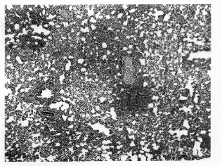
Figure 3.
Eosinopbilic abscess (MKbody) in
the liver of a ferret. The Splendore-Hoeppli
reaction at the center of the lesion
surrounds
a degenerating microfilaria
x2O0).
philic abscesses and granulomas, as observed in the fer-
ret.10 The prominence of lesions in the liver rather than lung
in the ferret may reflect a difference in the primary site of
microfilarial clearance, although pulmonary histopathology in
the ferret can be initially similar to that of TE in humans. As
in the TE syndrome, ferrets develop prominent immune re-
sponses to filarial antigens, including immediate hypersensi-
tivity to microfilarial antigens)11,12
Usefulness of Model
The ferret infected with
B. malayi
mimics several manifes-
tations of lymphatic filariasis in which knowledge of the
pathogenesis is incomplete. Persistent, severe filarial
Figure
4. Interstitialpneumonia in aferret
immediately after clearance of microfi-
lariae from the peripheral blood x 100).
lymphedema and the hyperresponsive syndrome of oc-
cult filariasis (TE) are not commonly reported in other ex-
perimental hosts, and the ferret will be of particular value
in study of these manifestations. As an experimental
model, the inflammatory reactivity and rapid development
of chronic lymphostatic disease parallels that of individu-
als migrating to endemic areas rather than natives to
these areas13; thus, the ferret model should have applica-
tion in examining the immunopathology of the acute syn-
dromes associated with infections in populations alien to
endemic areas. In addition, the high immune responsive-
ness to infection could be useful for evaluating hypersen-
sitivity reactions in treatment of lymphatic filariasis. The
ferret is increasingly used as an experimental animal in
|
|
3 |