|
2 |
|
diffuse interstitial pneumonia. The alveolar interstices are
infiltrated with histiocytes, eosinophils, and lymphocytes
that form occasional eosinophilic granulomas (Figure
4).2
Comparison With Human Disease
Lymphatic dwelling filariae induce a range of manifesta-
tions that include: 1) asymptomatic microfilaremia, 2) filar-
ial fevers with transient edema and recurrent adeno-
lymphangitis, 3) persistent lymphedema and elephantia-
sis, and 4) the tropical eosinophilia syndrome. It is
considered that the disease manifestations are immuno-
logically mediated and that asymptomatic, microfilaremic
infections are associated with a suppressed or restricted
immune reactivity.6 Ferrets infected with
B. malayi
have a
similar spectrum of clinical features. Asymptomatic micro-
filaremia is observed but most ferrets develop manifesta-
tions associated with high immune responsiveness. The
inflammatory reactions in the lymphatics and lymph
nodes parallel the lesions reported in humans.7 The acute
episodes of adenolymphangitis with fever, common in hu-
mans, are not obvious in the ferret but episodic lymph-
edema is present. Chronic lymphedema, a major medical
problem in lymphatic filariasis,8 is induced by multiple in-
fections in the ferret. Histopathologic changes are consis-
tent with filarial lymphedema, although the extreme der-
mal hyperplasia of elephantiasis has not been noted.
Tropical fitarial eosinophilia (TE) is considered to be a
hyperresponsive syndrome associated with immune
clearance of circulating microfilaria in the lung and other
organs. The hallmarks are amicrofilaremia, elevated IgE
levels, and high peripheral eosinophilia. The syndrome is
often expressed by respiratory signs resembling asthma
(tropical pulmonary eosinophilia).9 Early microscopic
changes of human pulmonary disease include histiocyte
infiltration of the interstitium that progresses to diffuse eo-
sinophilic pneumonitis. Occasional eosinophilic ab-
scesses are centered around microfilariae or their rem-
nants.9 Less attention has been paid to lesions in other
organs. In some patients, however, adenopathy and eo-
sinophilia may be the main presenting features. Hepato-
megaly and lymphadenopathy are especially common in
children and these organs usually contain microfilariae, in
the form of MK bodies, trapped within grossly visible eosino-
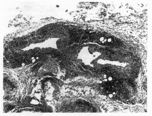
Figure 2.
Subcutaneous lymphatic vessel
with fibrotic walls and chronic inflamma-
tion (X100).
Figure
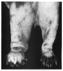
1.
Persistent edema of tbe right hind paw in a ferret af
ter multiple infections with
B. malayi
third-stage larvae.
|
|
2 |