
|
|
MetazoaThis group includes three subgroups (taxonomic vocabulary not clear here):{3940} 1. The acoelomates, which
includes the Phylum Platyhelminthes (and cestodes), which have no digestive tract. Cestodes
have calcareous corpuscles and a segmented body. 2. The pseudocoelomates, including Phylum Acanthocephala: no digestive tract, anterior end has proboscis Phylum Nematoda (although in other places the pseudocoelomate is the phylum and the others are classes): digestive tract present, no proboscis 3. The coelomates, including Phylum Pentastomida (Porocephalus, Armillifer, Linguatula): digestive tract present, no proboscis, cuticle with sclerotized openings, cephalic hooks, head and acidophilic glands Phylum Arthropoda: striated muscles, jointed appendages, chitin exoskeleton. Phylum Arthropoda contains two classes that contain important ectoparasites: Arachnida and Insecta. The arachnids all have reduced mouthparts and no head. Arachnid adults and nymphs all have 4 pairs of legs, whereas larvae have 3 pairs.{3936}
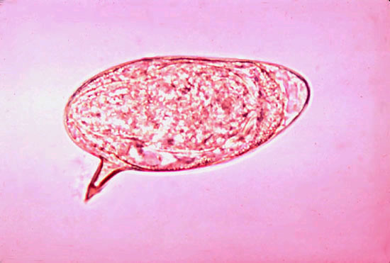
Phylum PlatyhelminthesTrematodes (flukes)These acoelomates have spongy bodies with no large cavities, and are not further divided into heads and bodies. The intestine is present but is bifurcated and ends blindly. The outside integument is a syncytial epithelium, often with spines. There are oral and ventral suckers. Just below the integument is an outer circular layer of muscle, middle longitudinal layer and inner diagonal layer. Muscle fibers may extend from dorsal to ventral in places. The adults are bisexual except the schistosomes. All eggs (except for schistosomes) are operculated and usually brown or yellow in color.{3940} Schistosoma
Fasciola hepaticaThis significant ruminant pathogen can be hosted by the rabbit as well, which may serve as a reservoir of infection in endemic areas. Laboratory rabbits are readily infected experimentally. Adults are found in the gall bladder and bile ducts. Metacercaria are encysted on vegetation and are the source of infection for the rabbit or ruminant. Pathology includes fibrous tracts in the liver, ulcers in the gall bladder, chronic cholangitis and hyperplasia of the bile ducts.{3936} Lab-acquired infections have occurred in people who contact aquarium snails, so Fasciola is listed as an ABSL2 organism.{3950} Dicrocelium dendriticumDicroceliosis is distributed worldwide in humans, sheep, goats, and cattle, causing (in the case of livestock) significant economic losses due to condemnation of the liver. Its life cycle makes laboratory study difficult: infective eggs develop in land snails, and metacercaria reach infective stages in ants (Formica). After ingestion by the definitive host, flukes migrate to the bile ducts via the portal vasculature. After some acute angiectasis of central veins, chronic inflammation of the bile ducts with marked fibrous tissue proliferation develops. Disease, if any, is caused by mechanical irritation caused by migrating flukes and toxicity of metabolic by-products. Three changes include hyperplasia of bile duct epithelium, peri-ductal inflammation, and fibrosis around a parasite egg that may be visualized using silver staining.{4086} Hamsters can be infected experimentally and are used to study liver changes induced by Dicrocelium. At 80-120 days the chronic stages have set in, and plasma ALT and AST are elevated, indicating active liver cell necrosis. Thiobarbituric acid-reactive substances (TBARS), markers of lipid peroxidation, are also elevated, as is the GSSG:GSH ratio (reduced: oxidized glutathione). These are indicators of oxidative stress, and may include generation of superoxide radicals. Bile flow paradoxically increases. The biliary tree contributes less bile, but the area around the canaliculus expands.{4086} Athesmia heterolecithoides (foxi)This is a common parasite of New World monkeys such as capuchins, squirrel monkeys, marmosets and titis, but there are no clinical signs and the flukes are easy to miss as they are quite small. They are found in the bile ducts of the monkey. The intermediate host is a mollusk.{4150}
Cestodes (tapeworms)Cestodes have at least two hosts, one in which the larva develops, and the other in which the adult develops. In the intermediate host, eggs are ingested, hatch in the intestine and are carried hematogenously to the liver, lungs, brain or elsewhere, where they develop into larvae. The definitive host ingests the intermediate host, in which the larvae grow to adulthood in the small intestine.{3935} Cestodes have dark-staining calcareous corpuscles in the parenchyma. There is no intestine. The larvae found in the intermediate host can form either cystic or solid structures. Those with solid structures are usually Spirometra, a rare parasite of dogs and cats which infects deer mice, voles, and man as intermediate hosts. The larvae encyst in connective tissues of the intermediate host, a condition called "sparganosis"{3935}. Cystic structures formed by cestodes are of five types{3940}:
In laboratory animals most cestodes are in the Order Cyclophyllidea. The scolex has 4 cup-shaped suckers, eggs are nonoperculated, the uterus is tubular or sacular, and the embryos are nonciliated and possess 3 pairs of small hooks. They have no respiratory or circulatory systems, and they lack an alimentary tract.{3936} An exception is Diphyllobothrium latum, which is a member of the Order Pseudophyllidea. This fish tapeworm has two intermediate hosts (a copepod and a fish) and the definitive hosts are dogs, cats and wild carnivores.{3935} Hymenolepis (was Rodentolepis)
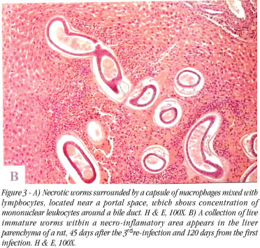
Human infection with H. nana has been reported, probably because of the heteroxenous lifestyle allowing either direct or indirect infections.{4141} It is therefore handled using ABSL2 procedures.{3950} H. diminuta is the rat tapeworm found in mice, rats, hamster, NHPs and man. Differentiation from H. nana is by the lack of hooks in the scolex, somewhat larger eggs without polar filaments, and the requirement for an intermediate invertebrate host.{3935} Hymenolepis microstoma is found in bile ducts of rodents and its eggs are larger than H. nana.{4756} Taenia taeniaformis/Cysticercus fasciolarisRats can serve as intermediate hosts for the cat tapeworm. Following ingestion of ova, cysts form in the liver. Host connective tissue capsules can develop into sarcomas, a potential carcinogenesis model.{2764} Taenia pisiformis/Cysticercus pisiformisThe rabbit can be an intermediate host for the dog tapeworm. Adult Taenia pisiformis are found in carnivores (usually dogs) which acquire infection by eating rabbit viscera. After 7 weeks the dogs pass gravid proglottids and embryonated eggs (hexacanth embryos or oncospheres) in the feces, which may contaminate field crops such as hay. In the rabbit, the oncospheres hatch, migrate via hepatic portal veins to the liver, where they mature to the cysticercus or bladderworm. They reach its surface in about 15 days, when they pass into the peritoneal cavity. Infective larvae may be found adhering to the mesentery or viscera. Younger metacestodes cause scarring and fibrosis as they migrate through the liver. Another dog tapeworm, T. serialis, has a more limited geographic distribution and the embryos migrate to inter-muscular connective tissues. A coenurus, a fluid-filled bladderworm 4-5 cm in diameter containing several scolices, is formed.{3936}{4145} A case report was published in 2001 concerning two Dutch Belted rabbits (from Myrtle's) which died, probably from unrelated causes, and at necropsy cysts were found in the abdomen. The cysts were 1 cm diameter, fluid-filled cysts with a distinct white spot (the scolex). They were either free-floating or attached to fat or omentum. Both rabbits' livers also had 2-5 cm scattered yellow shrunken areas and linear white streaks 2-4 mm in diameter. The cysts contained invaginated scolices with hooks and suckers but no ova, as well as calcaneous corpuscles, features of cestodes. The diagnosis was Cysticercus pisiformis. Rule-outs included Taenia serialis (Multiceps serialis) which forms a coenurus in muscles; Echinococcus granulosus which also forms a hydatid cyst found in the abdominal cavity; and Encephalitozoon cuniculi which causes granulomatous hepatitis. The source of infection in these two cases is unknown; the CSU facility had one other record of the condition in a laboratory rabbit.{4145} AnoplocephalidsThese tapeworms lack a rostellum and hooks, and the family is taxonomically complex. North American rabbits can be infected by Cittotaenia variabilis, Mosgovoyia pectinata americana, and Mosgovoyia perplexa. Adults live in the small intestine, with orbatid mites as the intermediate host. If large numbers of adults are present the rabbit may have catarrhal enteritis, intestinal perforation or obstruction. Good sanitation should be sufficient to protect laboratory rabbits.{3936} Prosthenorchis has been a problem in callitrichid NHPs; it is spread by the cockroach (Blatella germanica) and damages the terminal ileum, causing peritonitis, abscesses and sepsis. The adults are about 2cm long x 2mm diameter, and tan in color. {2765} RaillietinaSeveral species infect North American wild rabbits. Adults live in the small intestine and use ants as the intermediate host.{3936}
Phylum AcanthocephalaAcanthocephalans have hooks on their heads. The muscles look different than in nematodes, with small round muscles within the outer circular muscle layer. There is no intestine, and eggs are loose in the body cavity since there is no uterus. Grossly they resemble cestodes in that they are flat.{3940}
Phylum NematodaNHPs in particular are infected by a large number of different nematodes. A taxonomic system from the NHP literature is listed below.{3941} Subclass Secernentea (phasmidia) Order Rhabditida, superfamily Rhabditoidea (Strongyloides[ABSL2]) Order Strongylida
Order Oxyurida, superfamily Oxyuridea (Enterobius, Trypanoxyuris, Lemuricola). In rodents there are Aspiculuris and Syphacia. The rabbit pinworm is Passalurus ambiguus. The gerbil pinworm is Dentostomella translucida.{4543} Order Ascaridida: Trichosomoides in rats, Paraspidodera in guinea pigs
Subclass Adenophorea (aphasmids) Order Enoplida
StrongylesThe cuticle of strongyles has external longitudinal ridges in trichostrongyles. Third stage larvae have double lateral alae. The esophagus is club-shaped. The intestine has few multinucleated cells. There are three subgroups of strongyles:
Back to Metazoa Back to nematodes Top of page OesophagostomumThe most common nematode of Old World NHPs is Oesophagostomum (the nodular worm). Animals are infected through ingestion, and the larvae penetrate the large intestine producing nodules which rupture. Thiabendazole kills mature worms in the intestine, but not the larvae in the nodules.{2765} Back to Metazoa Back to nematodes Top of page TrichostrongylusSignificant species of trichostrongyles in the rabbit are: T. affinis: affects Sylvilagus floridanus and Lepus americanus in the US; adults found in cecum and large intestine; prepatent period 10-11 days; measure 5-10 mm long and males have spicules; used in studies of life cycle and host specificity T. retortaeformis: affects wild O. cuniculus, Lepus timidus in the UK and Australia; adults found in small intestine; prepatent period 10-11 days; measure 6-10mm long and males also have spicules; is capable of causing significant pathology and reducing populations Graphidium strigosum: found in domestic and wild O. cuniculus in the UK, Europe and Australia; adults found in stomach; prepatent period 12 days- 5 weeks; measure 8-20mm long, males have paired slender spicules; is capable of causing slight gastritis, severe infections may result in anemia{3936}. DictyocaulusThis parasite is the cause of verminous pneumonia in sheep and goats . Dictyocaulus filaria is the most pathogenic of the etiologic organisms, Muellerius capillaris is the most common and least pathogenic, and Protostrongylus rufescens is intermediate in pathogenicity. Dictyocaulus affects 2-18 month old sheep. They have chronic fever, cough, nasal discharge, tachypnea, anorexia and weight loss. Parasites may be found in the bronchi, especially the those of the diaphragmatic lobes. Pulmonary edema, emphysema, and atelectatic and pus-filled lobules are also sometimes found.{4512} Back to Metazoa Back to nematodes Top of page Obeliscoides cuniculiThe primary importance of this gastric nematode of rabbits is as a model for ruminant trichostrongyliasis. Studies of nutritional factors, arrested development of larvae in the stomach, and immune response have been performed. Both wild and domestic rabbits and hares are susceptible, ingesting infective third stage larvae. The larvae penetrate the stomach wall and develop into fourth stage and then adults. The adults are pinkish and are found in gastric mucus. Males are 10-15mm long and females are 15-18mm long.{3936} Back to Metazoa Back to nematodes Top of page Protostrongylus boughtoniThis metastrongyle infects wild rabbits (Sylvilagus is probably an abnormal host, but Lepus is a normal host) in North America. Like most metastrongyles, adults are found in the lungs where they cause bronchitis and peribronchitis. Eggs hatch to first stage larvae, are coughed up and swallowed and pass in the feces. Land snails (Vallonia pulchella) are the intermediate host, and the rabbit acquires infection by eating the snails. Larvae can be isolated by using the Baermann apparatus.{3936} Back to Metazoa Back to nematodes Top of page Oxyurids
Syphacia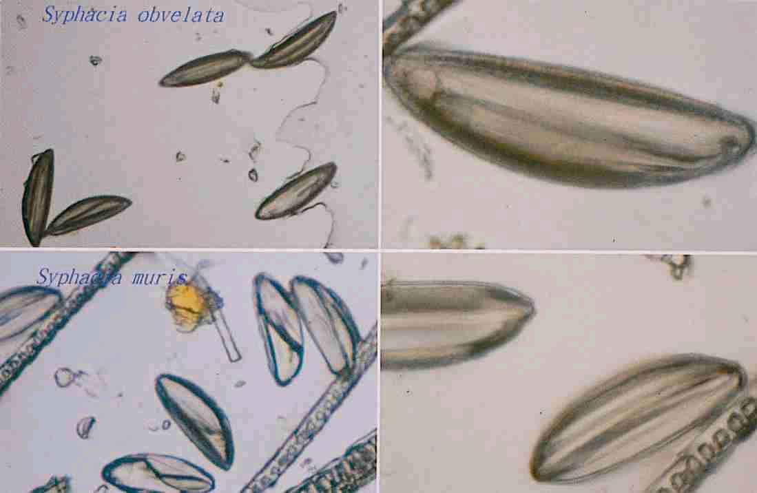
The mouse pinworm (S. obvelata) is found also in rats, hamsters and gerbils. S. muris infects the cecum and colon of the rat.{2764} There is great sexual dimorphism: adult female worms are 3.4-5.8mm, and the males are 1.1-1.5mm. Worms are found in the cecum and large intestine. In histologic sections, all pinworm adults are flat and have lateral alae{3940}. The life cycle is completed in 11-15 days, which is shorter than the life cycle of Aspiculuris tetraptera. Infection is more prevalent in mice 4-5 weeks of age, but it is asymptomatic. Eggs are deposited on the skin and hair of the perineum, so fecal flotation is not useful for diagnosis. Eggs are infective within a few hours and last for weeks in the environment. Diagnosis is by means of the tape test, or by finding adults in the intestine at necropsy. Treatment is by piperazine in the drinking water for 1 week, then wait a week and treat again during week 3. Other anthelmintics have also been recommended (mebendazole, trichlorphon, thiabendazole or dichlorvos){3551}.
Back to Metazoa Back to nematodes Top of page Aspiculuris tetraptera Back to Metazoa Back to nematodes Top of page
Passalurus ambiguus
Back to Metazoa Back to nematodes Top of page Dentostomella translucida
Ascarids (Order Ascaridida)Ascarids never have embryonated eggs, a method of differentiating them from spirurids. The eggs are thick shelled. Adults have a mouth with three large lips. Examples include Toxocara, Ascaris, Parascaris.{3940} There have been lab-acquired infections with Ascaris, Enterobius, and Strongyloides, as well as hookworms (which can cause an allergic response if aerosolized). These are handled using ABSL2 precautions.{3950} BaylisascarisRabbits are aberrant hosts for ascarids, and have been used as models for visceral larva migrans and cerebrospinal nematodiasis caused by B. procyonis (raccoon host) and B. columnaris (skunk host), as well as Toxocara canis, the dog ascarid. Eggs passed by the host are very resistant to the environment and don’t become infective for 30 days. After hatching, the larva migrate almost anywhere. If the CNS is infected, the rabbit develops torticollis, ear droop and loss of equilibrium. Granulomas are found in the brain. Histologic cross sections show two lateral alae.{3936} Back to Metazoa Back to nematodes Top of page Trichospirura (Superfamily Thelazoidea)
Trichosomoides crassicauda
Back to Metazoa Back to nematodes Top of page Paraspidodera uncinata
Back to Metazoa Back to nematodes Top of page
Filarids (Order Spirurida, Superfamily Filaroidea)The family Filaroididea has a rudimentary mouth with no lips. Males are usually much smaller than females with spicules that are unequal and dissimilar. Adults are found within body cavities, blood or lymphatic vessels, or connective tissues. Fully developed larvae (microfilaria) circulate in the bloodstream to be picked up by the required arthropod intermediate host, usually a mosquito.{3936} Filarid worms have a cuticle with ridges, bosses or annulations. Bulges of cuticle called lateral internal ridges are common. The intestine is very small, but these are the only nematodes with small intestines. Examples include Dirofilaria, Dipetalonema, Onchocerca, Stephanofilaria, and Brugia.{3940} DirofilariaRabbits are infested by either D. scapiceps (in Sylvilagus, Lepus and Oryctolagus), or D. uniformis (Sylvilagus in the southern US). Adults are found in the connective tissues of the hock and stifle (D. scapiceps) or the trunk (D. uniformis). D. scapiceps causes proliferative tenosynovitis. The adults have lateral alae and are spirally coiled, whereas D. uniformis adults have no alae and are straight. Both are common in rabbits housed outdoors and may be interesting as models of other filariid infections. Rabbits can also be aberrant hosts for the dog heartworm, D. immitis.{3936} Back to Metazoa Back to nematodes Top of page BrugiaB. malayi, found in the Far East, causes filariasis in man and animals. The only species found in the US are B. lepori from rabbits and B. beaveri from raccoons and bobcats. Adults are found in the subcutaneous tissues and abdominal lymphatics. They provide an interesting model for human filariasis; although zoonosis is not described it may be presumed to be possible because other members of the genus are zoonotic.{3936} Gerbils can be experimentally infected with B. pahangi and B. malayi{3560}. Cats have also served as models for lymphatic filariasis due to Brugia malayi. Advantages include the ability to determine the exact time of onset (humans in endemic areas are constantly exposed), the extent of infection with adult worms, and the effect of each parasitic stage on the lymphatic vessels.{4042} Lymphatic filariasis in the ferret (Fascicle) After injection of Brugia malayi into the footpad of the ferret, the animal may have three outcomes: asymptomatic microfilaremia, episodic lymphedema, or chronic lymphedema if infection is multiple. Chronic lymphedema is the most severe outcome in tropical lymphatic filariasis. The signs of elephantiasis in humans and ferrets are assumed to be caused by immune hyperresponsiveness. The lymphatics become blocked, and there is inflammation, fibrosis and edema. In the ferret, lesions are found more in the liver than in the lungs, as in humans; the liver may be the primary site of microfilarial clearance. The degenerating microfilaria are called Meyers-Kouwenaar or MK bodies, and they may be encased in eosinophilic refractile substance called Splendore-Hoeppli bodies. If necropsied during or after microfilarial clearance, ferrets have interstitial pneumonia. Back to Metazoa Back to nematodes Top of page Subclass Adenophorea (aphasmids), Order Enoplida,
Superfamily Trichuroidea
Trichuris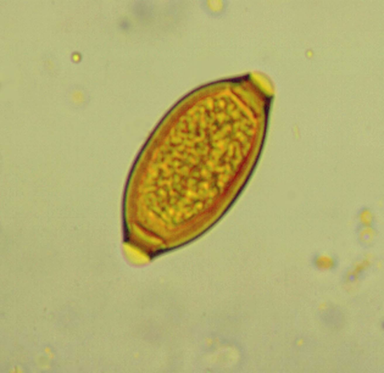
Rabbits are infected by both T. leporis (the whipworm of hares including Lepus and Citellus) and T. sylvilagi (infects Sylvilagus floridanus). Adults are found the large intestine and cecum and are larger than trichostrongyles, measuring 17-21 mm. Trichurid eggs are barrel-shaped, thick shelled and have plugs at each end.{3936}
Capillaria
In birds, capillariasis (hairworms) is rare in clean facilities. Species include C. obsignata (chicken, turkey and others), C. columbae (pigeons), and C. contorta (game birds). Signs include a sudden drop in egg production, unthrifty appearance and slight enteritis. Unlike C. hepatica, infection may spread via ingestion of 8-day embryonated eggs which develop in the crop and esophagus. Diagnosis is made by mucosal scrapings.{3568} Capillaria tamiasstriati is a parasite of the woodchuck small intestine.{3560} The nematode Capillaria
xenopodis (Pseudocapillaroides xenopi), a skin parasite of South
African clawed frogs (Xenopus laevis),
is quite common in laboratory animal facilities. It causes serious skin changes
and may further lead to wasting and death of affected frogs. Signs include lethargy, anorexia, skin color change,
rough “flaky” skin, decreased egg production in females, Other aphasmid parasites that primarily infect and cause hyperplasia of epidermal/epithelial surfaces include: Capillaria spp. (C. annulata, C. contorta, C. obsignata) in the crop and esophagus of birds; Capillaria philippinensis in the small intestines of humans; Capillaria bohmi in the nasal mucosa of canids; Trichosomoides crassicaudain the urinary bladder of rats; Anatrichosoma spp. of the skin and nasal mucosa of primates; and Gongylonema spp. in the nasal mucosa, oral mucosa, and esophagus of primates and cattle (from a AFIP Wednesday slide conference). Back to Metazoa Back to nematodes Top of page
Pentastomids (Phylum Pentastomida)Two families are of veterinary importance: Porocephalidae (Porocephalus and Armillifer) and Linguatulidae (Linguatula).{3936} Linguatula serrata adults live in the respiratory tract of the dog, fox, wolf, human, goat and sheep. Rabbits ingest the eggs expelled from the host, which develop into larvae. Larvae puncture the bowel wall and develop into infective nymphs in the mesenteric lymph nodes. Infestations in rabbits are an incidental finding at necropsy{3936}. Porocephalus clavatus nymphs adhere to the surface of organs in the abdominal and thoracic cavities of callitrichid NHPs; they are considered an incidental finding.{2765} The parasites are often C-shaped and found in the peritoneum of NHPs. The cuticle has sclerotized openings which stain black with movat, a pentochrome stain. They have a mouth, digestive system and anus. This is the only animal with eosinophilic glands surrounding the alimentary canal. The 4 hooks of the mouth attach to the lung. Adults parasitize the respiratory system of amphibians, reptiles and some birds.{3940} Snakes can become infected with Armillifer by ingesting monkeys; the intermediate monkey hosts are infected by eating things contaminated with snake feces containing eggs. The larvae are found in the NHP peritoneum. (CL Davis 2001)
Phylum ArthropodaClass ArachnidaLicePolyplax serrata
Gliricola porcelli
Columbicola columbae
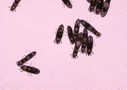
This is the slender pigeon louse, which is long and slender (2½mm long x ½mm wide) with prominent claws on its legs. It is also a Mallophagan, having chewing mouthparts and yellow in color. The mesothorax and metathorax are fused into one piece. The tarsi of species parasitic for birds have 2 claws, and those parasitic on mammals have one claw. Filiform antennae are visible at the sides of the head. Mallophagans spend their entire life on the host. Eggs are laid in clusters on feather shafts and hatch in 4-7 days. Nymphs mature in 3 weeks. Adults live for several months. They cause feather abrasion. Lice are transmitted by direct contact or by the pigeon ked, Pseudolynchia canariensis.{4097} Goniocotes gallinae
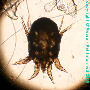
The fluff louse of chickens and guinea fowl is also a Mallophagan. It is pale yellow and nearly circular, 1.5mm long. The life cycle is unknown, but it is presumed to spread by direct contact. It generally causes little irritation aside from restlessness and damaged feathers. Topical treatment with permethrin dust eliminates Mallophagans.{4097} TicksTick-borne diseases, which are emerging as public health threats at the rate of about 1 per decade, include Lyme disease (Borrelia burgdorferi), human granulocytic ehrlichiosis (HGE, Ehrlichia phagocytophila), Rocky Mountain spotted fever (Rickettsia rickettsii), and canine ehrlichiosis (E. canis and others). Bartonellosis (in dogs, Bartonella vinsonii) is suspected of being tick-borne. Brown dog ticks (Rhipicephalus sanguineus) are the vector of E. canis. The lone star tick (Amblyomma americanum) is the suspected vector of B. vinsonii. The deer tick (Ixodes scapularis) lives in the NE and upper Midwest US and is the principal vector of Lyme disease, human babesiosis, and HGE. One study detected a correlation between the presence of deer ticks and seropositivity to B. burgdorferi and the HGE agent in dogs in Rhode Island.{4580} MitesIt is difficult to diagnose mite infestations on physical exam, so some ancillary techniques are useful. Examination with a dissecting microscope in anesthetized or dead animals, pressing tape on the pelage of recently-killed animals, or placing the dead animal on black paper in a rectangle of double-sided tape for 18 hours will pick up the mites as they leave the cooling carcass. Skin scrapings will miss both fast movers and low numbers of slow movers; this technique is useful for Demodex, Notoedres and Sarcoptes, which are not found on laboratory mice.{3551} Ectoparasitism can be extremely difficult to eradicate in a rodent colony; periodic control methods are usually used unless gnotobiotic rederivation is possible. The easiest treatment (perhaps somewhat outmoded in the third millennium) is taping dichlorvos strips in the cages; the mice will have a transient decrease in acetylcholinesterase.{3551} Ornithonyssus bacoti
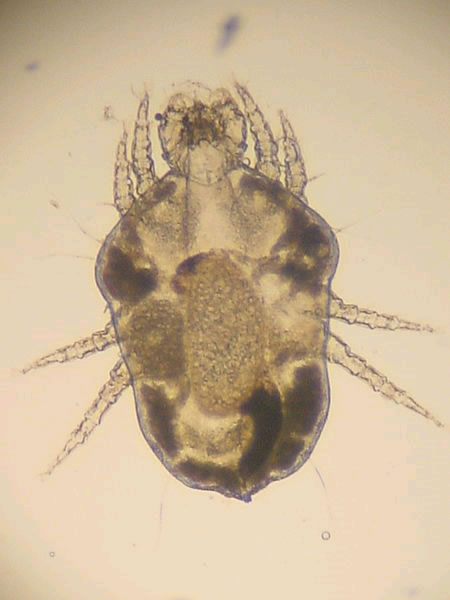
Myocoptes musculinis
The mouse mite is the most common ectoparasite of mice. Note the "cups" on the ends of the legs. Eggs are attached to the middle third of the hair shaft. The mites inhabit larger areas of the body than Myobia, found in the inguinal region, abdomen, back, head and neck. It feeds on surface epithelium. It belongs to the Astigmata along with the sarcoptic mites, in which there is marked sexual dimorphism. It resembles Trichoecius romboutsi. Signs are patchy alopecia with erythema, but not usually pruritis.{3551}{3935} However, a substantial immunologic disorder can occur in BALB/c mice infested with Myocoptes musculinis, termed mite-associated ulcerative dermatitis (MAUD). BALB/c mice are resistant to dermatitis induced by Myobia musculi.{4096} Myobia musculi, Radfordia affinisThe common fur mite of the mouse is found on the back, neck and shoulders. It is usually subclinical, but hypersensitivity can occur (especially in C57BL) with ulcerative dermatitis, pruritis, and erosion of the pinnae. Mice are usually co-infected with several mite species. In rats, the mite is Radfordia ensifera{2764}. Myobia is a surface dweller, feeding on skin secretions, and does not invade deep into the skin at any phase of its life cycle. Transmission requires close direct contact. Atopic dermatitis induced by Myobia has been studied since the 1970s. NC/Nga mice were developed in Japan based on Japanese fancy mice in 1957. NC/Jic mice were derived from NC/Nga. They were originally recognized as having autoimmune disease, the most evident of which was dermatitis, and are considered a model of human atopic dermatitis. Signs develop on the face, neck, ears, and dorsal skin beginning at 6-8 weeks of age. Plasma IgE is elevated. Severity and age of onset depend on conditions in the animal facility, although it cannot be eradicated by sanitation alone. Clinical signs wax and wane, and there may be reduced reproduction efficiency. It was observed in 2000 that although conventional mice developed dermatitis, mice housed in SPF conditions did not. It was shown that dermatitis was associated with mite infestation, and could be transmitted by co-housing with non-infested mice. Cellophane tape testing of the fur on the head, neck and shoulders was used to document infestation. Treatment was with ivermectin (0.1mg/ml) diluted 1:100 and delivered as 4 squirts per cage during the weekly cage change (0.26mg ivermectin) and continued for two weeks after eradication. Dermatitis scores declined significantly with treatment.{4096} Myobia (left) has a single empodial claw on the second pair of legs; Radfordia (right) has a double empodial claw. Note that the first pair of legs surrounds the mouthpart area.{3763}
Sarcoptes scabei
Adult female mites burrow in the skin to deposit ova. Mites develop to maturity in 17 days after hatching. Larvae and nymphs on the surface of the skin are responsible for transmission to other hosts. The burrowing mites, feeding on epithelial cells and lymph, cause intense pruritis with subsequent alopecia and serous exudate. Over time, a whitish-yellow crust of serum and debris develops. Lesions first appear on the nose and lips, extending to the eyes, forehead, face and occasionally external genitalia. Mange can be severe, including anemia and leukopenia, leading to debility and death in a few weeks.{3936} Diagnosis is made on deep skin scrapings cleared with 10% KOH. Adults are small and round with short legs, the rear two pair of which do not extend beyond the edge of the body. Pedicels are long and unjointed, some with bell-shaped suckers (legs 1&2 in females, 4 in males).{3936} Trixacarus caviae
Psoroptes cuniculi
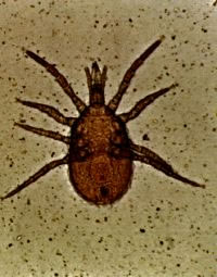
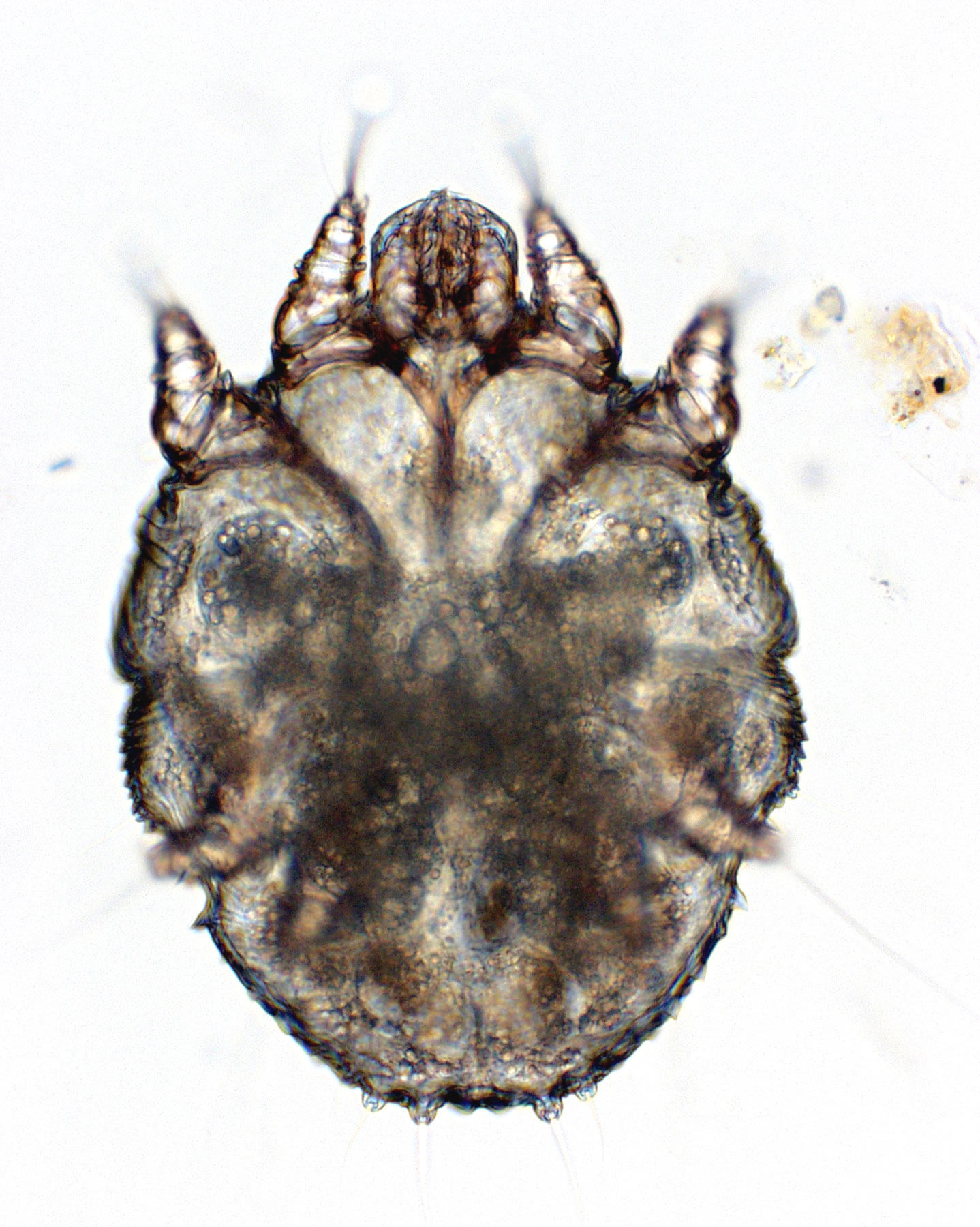
This mite causes otoacariasis (ear mange, ear canker) in rabbits. There may be cross-infestation between various animal species with it. Its incidence is, surprisingly, relatively high in captive rabbits but unknown in wild lagomorphs. Psoroptes are found exclusively on the inner surface of the pinna, where they do not burrow deeply. All stages of the life cycle (21 days) are found on the host. The lesions (hypertrophy of the Malpighian layer, parakeratosis of the horny layer, and epithelial sloughing) may be the result of an allergic response. Adults can be seen with the unaided eye. They have legs that project beyond the margin of the body, and their pedicels are jointed. Infested rabbits can be treated with ivermectin (400-440m g/kg SQ, given twice at 1-2 week intervals). Mineral oil, alone or in combination with topical acaricides, has been used successfully for years.{3936} Cheyletiella
Psorergates simplexThis mouse mite inhabits hair follicles, causing dermal cysts that are visible from the underside of the skin at necropsy. The cysts contain mites and debris. It has not been reported from well-managed colonies for a decade or more{3551}. It has been reported in meadow voles{3560}. DemodexIn hamsters, two species (D. aurati and D. cricetus) are common. Clinical signs are uncommon. Alopecia on the rump may occur, with dry scaly skin. Diagnosis is made by skin scraping.{3566} In mice, the serendipitous finding of Demodex musculi was reported in 1999 in a few strains of immunocompromised mice. The mites were very superficial, being found on plucked hair samples, and when seen histologically in the hair follicles appeared to cause no inflammation. Demodex musculi is shorter than other members of the genus: females 113-170um, males 126-154um.{4569} Chirodiscoides caviae
Leporacarus gibbus
Order DipteraFamily Hippoboscidae (louse flies and keds)Diptera have functional mesothoracic wings and a pair of club-shaped balancing organs on the metathorax called halteres. Members of the family Hippoboscidae lack halteres and wings, or they have only temporary wings. They are relatively permanent ectoparasites of birds and some mammals.{4097}
|
|
©1999, Janet Becker Rodgers, DVM, MS, DipACLAM, MRCVS All rights reserved. Comments? Send an email to janet.rodgers@vet.ox.ac.uk |
 This parasite causes serious disease in humans but is an incidental finding
at necropsy in NHPs. Species include S. mansoni (squirrel monkeys,
several Old World species, and great apes), S. haematobium (Old World
monkeys and apes), and S. mattheei (baboons). Since the intermediate
host is a snail, there is little danger of zoonotic spread. However,
lab-acquired infections have occurred in people who contact aquarium snails,
so the organism is listed as an
This parasite causes serious disease in humans but is an incidental finding
at necropsy in NHPs. Species include S. mansoni (squirrel monkeys,
several Old World species, and great apes), S. haematobium (Old World
monkeys and apes), and S. mattheei (baboons). Since the intermediate
host is a snail, there is little danger of zoonotic spread. However,
lab-acquired infections have occurred in people who contact aquarium snails,
so the organism is listed as an  H. nana
(the dwarf tapeworm) is a zoonotic tapeworm found in the small intestine of
rodents
(mice, rats, hamsters, fat sand rats), NHPs (rhesus, squirrel monkey and chimpanzee) and
man.
Alternative name may beVampirolepis nana{
H. nana
(the dwarf tapeworm) is a zoonotic tapeworm found in the small intestine of
rodents
(mice, rats, hamsters, fat sand rats), NHPs (rhesus, squirrel monkey and chimpanzee) and
man.
Alternative name may beVampirolepis nana{
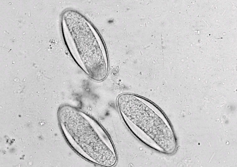 Female pinworms in the mouse are slightly shorter than S. obvelata;
otherwise the best way to make the distinction is to examine the eggs. Aspiculuris
eggs are symmetrical, whereas Syphacia eggs are asymmetrical{
Female pinworms in the mouse are slightly shorter than S. obvelata;
otherwise the best way to make the distinction is to examine the eggs. Aspiculuris
eggs are symmetrical, whereas Syphacia eggs are asymmetrical{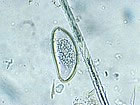 The
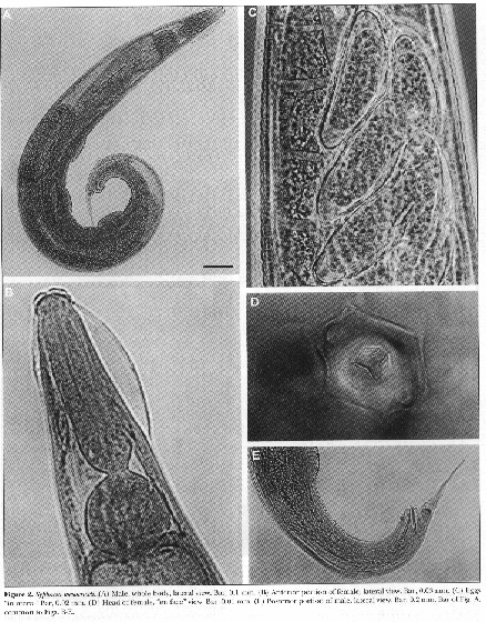
rabbit pinworm is found in both wild and domesticated rabbits in the US
and elsewhere. Juvenile stages are found in the small intestine and cecum.
Adults are found in the large intestine and anterior cecum, and are grossly
visible as 4-8 mm long worms. The female’s tail tapers and has about 40
circular cuticular striations that look like a corkscrew. The eggs are flattened on one side. Piperazine,
phenothiazine, fenbendazole and probably ivermectin are curative.{
The
rabbit pinworm is found in both wild and domesticated rabbits in the US
and elsewhere. Juvenile stages are found in the small intestine and cecum.
Adults are found in the large intestine and anterior cecum, and are grossly
visible as 4-8 mm long worms. The female’s tail tapers and has about 40
circular cuticular striations that look like a corkscrew. The eggs are flattened on one side. Piperazine,
phenothiazine, fenbendazole and probably ivermectin are curative.{ This
pinworm usually infects Mongolian gerbils (Meriones unguiculatus) but can
also infect golden hamsters (Mesocricetus auratus) and great gerbils (Rhombomys
opimus). It lives in the small intestine and even the stomach, so fecal
flotation is necessary for detection. Eggs are passed only intermittently, a
source of false-negative results. The prepatent period is longer for this
pinworm than for others, around 25-29 days. The preferred treatment is
fenbendazole in the feed (150ppm) given every other week for 9 weeks total.
Ivermectin and piperazine, which paralyze the parasites, may not work as well
because of the distance the parasite must travel during expulsion.{
This
pinworm usually infects Mongolian gerbils (Meriones unguiculatus) but can
also infect golden hamsters (Mesocricetus auratus) and great gerbils (Rhombomys
opimus). It lives in the small intestine and even the stomach, so fecal
flotation is necessary for detection. Eggs are passed only intermittently, a
source of false-negative results. The prepatent period is longer for this
pinworm than for others, around 25-29 days. The preferred treatment is
fenbendazole in the feed (150ppm) given every other week for 9 weeks total.
Ivermectin and piperazine, which paralyze the parasites, may not work as well
because of the distance the parasite must travel during expulsion.{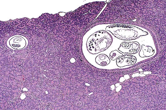

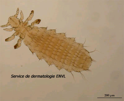 The common louse of laboratory mice is a sucking louse that causes
anemia
and is the
The common louse of laboratory mice is a sucking louse that causes
anemia
and is the 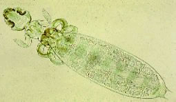 Biting lice (Mallophagans) have strong jaws and a free thoracic segment that
is narrower than the head. G. porcelli is yellow and measures 1-1 ½ mm
long, readily seen without magnification. Mild infestations cause no clinical
signs, and may not be diagnosed until after death, when the lice migrate to the
ends of the hairs.{
Biting lice (Mallophagans) have strong jaws and a free thoracic segment that
is narrower than the head. G. porcelli is yellow and measures 1-1 ½ mm
long, readily seen without magnification. Mild infestations cause no clinical
signs, and may not be diagnosed until after death, when the lice migrate to the
ends of the hairs.{
 This mite has been found on mice,
rats, hamsters, cats, wild carnivores, chickens and other birds, and man. It
often gains access via wild rodents and hides in crevices. Mites are on the
host only to feed, every 2-3 days. Eggs are laid in crevices off the host,
hatching in 1-4 days. The entire life cycle is completed in 13 days or more.
The mite is a blood feeder, causing anemia, decreased reproduction and
death. It
transmits Rickettsia typhi,
This mite has been found on mice,
rats, hamsters, cats, wild carnivores, chickens and other birds, and man. It
often gains access via wild rodents and hides in crevices. Mites are on the
host only to feed, every 2-3 days. Eggs are laid in crevices off the host,
hatching in 1-4 days. The entire life cycle is completed in 13 days or more.
The mite is a blood feeder, causing anemia, decreased reproduction and
death. It
transmits Rickettsia typhi, 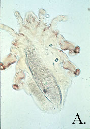

 Several species of Cheyletiella
infest animals. C. parasitovorax, C. takahasii, C. ochotonae, and C.
johnsoni are rabbit parasites. C. yasguri infests dogs, and C.
blakei infests cats. Although all stages of the life cycle occur on the
host, they may "bite and run" on humans and adults are rarely
identified even on heavily-infested animals. Cheyletid mites live on the skin
surface, found on the dorsum of the trunk near the scapulae. Eggs are found
attached to hair shafts 2-3mm above the base. Diagnosis is by examination of
brushings from the skin, under a dissecting microscope. The most distinguishing
feature of the adults are the C-shaped palpal claws. Infested rabbits can be
treated with silica gel products, topical acaricides, or ivermectin.{
Several species of Cheyletiella
infest animals. C. parasitovorax, C. takahasii, C. ochotonae, and C.
johnsoni are rabbit parasites. C. yasguri infests dogs, and C.
blakei infests cats. Although all stages of the life cycle occur on the
host, they may "bite and run" on humans and adults are rarely
identified even on heavily-infested animals. Cheyletid mites live on the skin
surface, found on the dorsum of the trunk near the scapulae. Eggs are found
attached to hair shafts 2-3mm above the base. Diagnosis is by examination of
brushings from the skin, under a dissecting microscope. The most distinguishing
feature of the adults are the C-shaped palpal claws. Infested rabbits can be
treated with silica gel products, topical acaricides, or ivermectin.{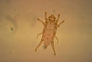
 The
rabbit fur mite does not cause overt disease and is difficult to detect. Mites
are found on the back and abdomen. Adults have a trapezoidal dorsal projection
extending over the mouthparts. Dense chitin gives the adults a brown
appearance. There is sexual dimorphism in that the males have large clasping
organs at the posterior end and are smaller than females. Combing or brushing
the hair and examining the hair under a dissecting scope is the means of
diagnosis. Infestation of laboratory rabbits was reported in CT, with the
finding of dark specks on fur under the tail.{
The
rabbit fur mite does not cause overt disease and is difficult to detect. Mites
are found on the back and abdomen. Adults have a trapezoidal dorsal projection
extending over the mouthparts. Dense chitin gives the adults a brown
appearance. There is sexual dimorphism in that the males have large clasping
organs at the posterior end and are smaller than females. Combing or brushing
the hair and examining the hair under a dissecting scope is the means of
diagnosis. Infestation of laboratory rabbits was reported in CT, with the
finding of dark specks on fur under the tail.{