
|
|
Fungal diseasesRingworm [ABSL2] Blastomyces [ABSL2] Coccidioides [ABSL2]
Histoplasma RingwormMice and rats are very rarely affected by Trichophyton mentagrophytes. If they are affected they develop alopecia and focal crusts on the head.{3763} Rabbits may be similarly affected by Trichophyton mentagrophytes, Microsporum canis or Microsporum gypseum.{3768} In neonatal guinea pigs, T. mentagrophytes is fatal in almost all animals. Culling is advisable as sows usually have recurrent infections at parturition.{3775} Young ruminants are often infected with T. mentagrophytes, particularly around the eyes and face.{4206} Aspergillus fumigatusLesions are occasionally found in rabbits at necropsy, and are nodules with a central area of necrosis surrounded by mononuclear cells and giant cells. Fungal hyphae can be demonstrated with silver stain or PAS.{3768} Blastomyces dermatitidis
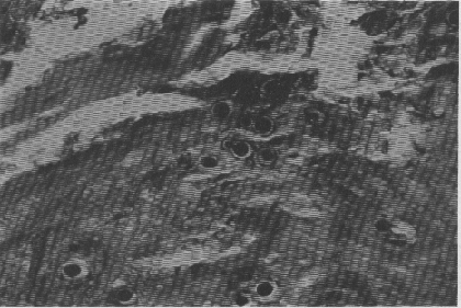
The eastern and midwestern US are the most common places to acquire blastomycosis. This is a dimorphic fungus which affects the lungs, skin and bone. Lesions are granulomas and suppurative nodules with caseous necrosis. It affects humans and dogs mostly. Finding organisms in tissue sections supports the identification of the fungus. Yeasts are large (5-20m), thick-walled and spherical. The cell wall does not stain. Buds are attached to mother cells by a wide base. In the photograph, organisms in the skin of a dog are seen surrounded by a clear zone which is the non-staining (on PAS) cell wall.{3953} It is handled using ABSL2 techniques. Candida albicans
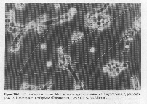
Candida is a ubiquitous organism causing opportunistic infections in immunocompromised or antibiotic-treated animals such as NHPs. Signs are dysphagia and a white pseudomembrane in the mouth. The lesion rarely penetrates past the basement membrane. H&E stain reveals organisms, but PAS or GMS stains are better{3770}. The photograph shows a dark-phase illumination of septate hyphae and oval budding blastospores from Carter{3953}. Coccidioides immitis
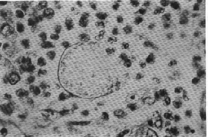
Unlike histoplasmosis, coccidioidomycosis occurs in arid regions, for example in the southwest US. It is perhaps the most infectious fungal disease. Disease occurs in many species of large animals and is usually confined to the lungs. Disseminated disease occurs depending upon the dose of pathogen inhaled and the host’s immune status. The meninges, joints, bones and subcutaneous tissues are affected by systemic disease. The lesions are suppurative and granulomatous. The photograph shows a ruptured spherule (very large, 10-80m) that has released endospores (2-5m) with inflammatory cells surrounding the area. Infection usually results in strong immunity, and serologic tests are more useful for this disease than for the others. There is a coccidioidin skin test as well as precipitin, complement fixation, immunodiffusion and latex agglutination tests. Kits of tests for multiple fungal diseases are available.{3953} Soil thought to contain Coccidioides and cultures must be handled using BSL3 precautions, since the arthroconidia are so tiny. Animal studies in which the route of challenge is parenteral are conducted at ABSL2. Cryptococcus neoformansThis organism is a yeast that primarily affects the CNS and eye, as well as the respiratory system. It has a very thick mucinous capsule that is obvious on India ink preparations. In dogs and cats, infection begins in the paranasal sinuses and extends later to the brain and meninges, or to the lungs. Ferrets, guinea pigs and birds have also been infected, as have a few NHPs.{3953} In NHPs, the meningeal lesions are gelatinous nodules or cysts with sparse granulomatous inflammation. There are abundant organisms demonstrable in these lesions.{3770} This is an animal biosafety level 2 organism. Histoplasma capsulatum
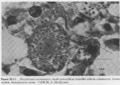
This is a dimorphic fungus that exists as a yeast at body temperature and a fungus at cooler temperatures. It grows best in soil with a high nitrogen content, explaining why it is found in areas contaminated with bird or bat feces. Almost all infections are asymptomatic, with inhaled organisms killed or encased in granulomas. Host immunity and the number of inhaled conidia may determine whether disease develops. If it does, it is a facultative intracellular parasite of macrophages of the reticuloendothelial system. Ulcers and tubercles are the main lesions. Humans and dogs are most susceptible but disease has been reported in other large animal species including NHPs. It can be difficult to see in smears because of its size; use oil immersion and examine monocytes, as in the photo. Diagnosis is by immunodiffusion combined with complement fixation to determine the presence of active infection.{3953} It is an ABSL2 agent, with several human cases occurring in diagnostic lab settings. The conidia are <5µ and are aerosolized and inhaled easily.{3950} An as-yet unclassified yeast organism was seen in three adult non-geriatric wild-caught Peruvian owl monkeys. Multiple yeast cultures on Sabouraud Dextrose Agar, SABHI Agar with blood, and SABHI Agar with Chloramphenicol and Cycloheximine were negative. However, paraffin-embedded formalin-fixed stained tissues (spleen, liver, kidneys, lymph nodes, heart, lungs, adrenal glands, and bone marrow) demonstrated myriad spherical to oval, thick-walled, uninucleate yeast cells 8-10 microns in diameter. A few cells had narrow-based budding of single daughter cells. Chains of organisms were not formed (as in Loboa). Yeast cells were common both free in the tissue and inside macrophages, but inflammatory infiltrates were absent. The yeast cell morphology was consistent with Histoplasma capsulatum var duboisii. The yeast cells tested from one animal were positive by FA testing to H. capsulatum and Blastomyces dermatitidis using a bivalent conjugate; however, the serum tested from this animal was negative by immunodiffusion and complement fixation for antibodies to both yeast species. One other monkey had yeast cells detected in his bone marrow. The Gridley Fungus Stain imparted a magenta color to the cell wall against the yellow counter stain, enhancing visibility of the yeast cells.{3619} Although its morphology was most consistent with Histoplasma capsulatum var duboisii, the absence of cellular reaction, inability to grow the organism on artificial media and the fact that the monkeys were from South America (rather than Africa) are inconsistent with that organism. Blastomyces dermatitidis have multiple nuclei and single broad-based buds and evoke pyogranulomatous inflammation, but grow on artificial media. Cryptococcus neoformans causes minimal cellular response like this organism, but it has a carminophilic capsule and is also readily cultivated. Loboa loboi can't be grown artificially, but is known only in humans and dolphins; it also infects only the skin, sub Q and lymph nodes, not viscera, evokes inflammation and has a characteristic chain pattern of budding.{3619} |
|
©1999, Janet Becker Rodgers, DVM, MS All rights reserved. Comments? Send an email to rodgers@uky.edu |